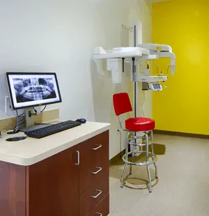
In order to create an ideal and detailed treatment plan, Dr. Amin and Dr. Vaziri need to have the best understanding of the position of your teeth within the bone and the position of the jaws within the skull. Not having the proper X-ray images and trying to come up with a treatment plan is like being in a dark room and trying to find the way out!
During your consultation appointment, some X-ray images will be taken to provide the necessary information for your customized treatment plan.
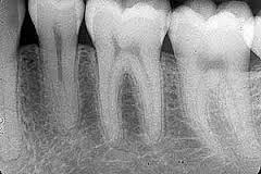
- Small, high resolution, two dimensional image
- Focused on a limited area, one or two adjacent teeth and their surrounding bone structure
- Orthodontists usually use it to evaluate the bone level supporting the teeth, to detect any abnormality on the roots of the teeth or for the placement of temporary anchorage devices
- A family dentist usually uses it to diagnose cavities and to provide root canal therapy
- Minimal exposure to radiation
The disadvantages are these images are two dimensional and too many images are required to cover the whole mouth.
- One of the most common radiographs taken in an orthodontic office
- Covers a broad area including both jaws, chin, all teeth, maxillary sinuses and part of the neck and the nose in one image
- Excellent tool for Dr. Amin Movahhedian and Dr. Hamed Vaziri to evaluate teeth development, chance of crowding or spacing before happening, finding out if there are any missing, extra or impacted teeth in the bone, and position of the wisdom teeth related to other teeth
- Minimal exposure to radiation, about the same as four periapical images
The disadvantages are these images are two dimensional, images of the different structures may overlap each other, images of the objects in the picture may not be proportionate to their actual size, and the lower resolution makes it a poor tool for diagnosing cavities.
- It is a radiograph of the profile of a patient
- Commonly used by orthodontists to evaluate the dental and skeletal relationship of upper and lower teeth and jaws. For example, for patients with severe overjet, also known as overbite, this X-ray will help the orthodontist to diagnose if the problem is the prominent upper jaw or a weak lower jaw. The cause will significantly affect the treatment approach
Disadvantages here are images of different structures and the opposite side may overlap each other, images and the actual profile may not be exactly the same size, only one side of the face is represented, and the images are two dimensional.
Cone Beam Computed Tomography (CBCT)
As mentioned above, the main disadvantage of the previous X-rays is that two-dimensional images are representing a three-dimensional structure. CBCT, on the other hand, provides a three-dimensional representation which makes it one of the best tools for precise diagnosis and treatment planning. Disadvantages here are increased radiation and a higher cost than regular X-rays.
- All of the structures are the actual size and there is no magnification
- There is no overlapping of the image
- It provides valuable detailed information about the position of the impacted tooth within the bone, and to detect if the adjacent teeth are damaged by it. It can also be used as a communication tool between the orthodontist and oral surgeon in terms of treatment planning for surgical orthodontics
- Implant dentistry has been revolutionized by the details that CBCT can provide about the density of the bone and where to place the implant
Computer Aided Surgical Stimulation
A fairly new approach that uses the data from CBCT to virtually plan the surgical orthodontic cases. This approach provides the most comprehensive treatment planning that is available today. We would like you to know that we are one of a few offices in the nation that utilizes this technique.
child x-rays in league city, orthodontics images, ava orthodontics league city, imaging orthodontics, human teeth length, league city child x-rays, cypress imaging, fry orthodontic specialists, dr vaziri dentist, images orthodontics, true image orthodontics, x ray machines cypress tx, true image orthodontics cypress, av imaging, orthodontic images, lateral cephalometric imaging, ava orthodontics, paño fin, lateral ceph x ray cost, image orthodontic, imagine orthodontics, fry orthodontic, ava orthodontics cypress, ava orthodontics spring, league city child x rays, child x rays in league city, root canal treatment league city, root canal league city, orthodontic imaging johnson city, orthodontic evaluation johnson city, league city root canal, braces for teenagers johnson city, advanced orthodontic treatment johnson city, orthodontic x rays johnson city, living with braces johnson city, orthodontic assessment johnson city
child x-rays in league city, orthodontics images, ava orthodontics league city, imaging orthodontics, human teeth length, league city child x-rays, cypress imaging, fry orthodontic specialists, dr vaziri dentist, images orthodontics, true image orthodontics, x ray machines cypress tx, true image orthodontics cypress, av imaging, orthodontic images, lateral cephalometric imaging, ava orthodontics, paño fin, lateral ceph x ray cost, image orthodontic, imagine orthodontics, fry orthodontic, ava orthodontics cypress, ava orthodontics spring, league city child x rays, child x rays in league city, root canal treatment league city, root canal league city, orthodontic imaging johnson city, orthodontic evaluation johnson city, league city root canal, braces for teenagers johnson city, advanced orthodontic treatment johnson city, orthodontic x rays johnson city, living with braces johnson city, orthodontic assessment johnson city


